Who we are

Was founded in 1987 by professionals with extensive experience, who were pioneers of Nuclear Medicine in Portugal.

The practice is installed in an area of 500 m2, designed specifically for Nuclear Medicine. Atomedical has three tomographic Gamma Cameras with two detectors. One (GE OPTIMA 640) equipped with a CT system for attenuation correction and to fuse anatomical with functional images (SPECT/CT).

The computer image processing systems consist of 4 workstations with networked software and are fitted with the latest methodological developments. All examinations are archived in a specific system (PACS). The marking and handling of radiopharmaceuticals is performed on shielded chambers with sterile conditions in accordance with regulations.

For radiation protection, the environment is constantly monitored by an automatic system, ensuring efficient protection of patients, employees Laboratory and the surrounding area. Atomedical has emergency systems (UPS). In case of power supply interruption, UPS guarantee the test continuity, which is an important problem in nuclear medicine for radiation protection of the patient. All software quality assurance and maintenance are carried out by the technical services contracted with the equipment suppliers and the laboratory technicians.

In Atomedical, our goal is not just to implement last generation technological developments, all our professionals follow these developments through proper training.

Team

Guilhermina Cantinho

Nuclear Medicine Specialist since 1986;

Clinical Director of Atomedical since 1987;
Nuclear Medicine MD and teacher at the Nuclear Medicine Institute of the Lisbon Medical School from 1987 to 2009.
Director of the Institute of Nuclear Medicine, Lisbon Medicine School from 2009 to 06/2022;
Invited Professor at the Lisbon Medicine School and Chair Regent of Nuclear Medicine at Lisbon Medicine School from 2009 to 06/2022.

Helena Pena

Nuclear Medicine Specialist since 1999;
Nuclear Medicine MD and teacher at the Nuclear Medicine Institute of the Lisbon Medical School from 1999 to 08/2022.

Arminda Veiga

Cardiology Specialist since 1997;
Graduated Hospital Assistant (Hospital de Santa Maria - Centro Hospitalar Universitário Lisboa Norte).

José Monteiro

Cardiology Specialist since 1992;
Graduated Hospital Assistant.

Afonso Nunes Ferreira

Cardiology Specialist since 2021;
Hospital Assistant (Hospital de Santa Maria-Centro Hospitalar Universitário Lisboa Norte).

Fernando Godinho

Physicist since 1963
;
Ph.D. in Biophysics, Emeritus University Professor Atomedical's Responsible for Radiation Protection and Quality Control.

Teresa Rézio

Medical Physics Specialist

Lurdes Gano

Doutorada em Farmácia;
Investigadora do Grupo de Ciências Radiofarmacêuticas do Instituto Superior Técnico; Responsável pela radiofarmácia na Atomedical;

/ Nuclear Medicine Tech.

Deolinda Cerqueira
Marco Pinheiro
Alexandra Alves
Alexandra Santos
Beatriz Lima
Cristiana Subtil

/ Secretariat

Alice Silva
Sandra Santos
Helena Mendes
Sara Martinho
Liliana Martins
Neuza Marques
Sandra Godinho

Close

Exams

Scintigraphy and SPECT/CT

Faq

What is a Scintigraphy?

A Scintigraphy is a functional imaging of an organ, useful for your doctor to evaluate the disturbance degree of that organ, to complete a diagnosis and thus contributing to decide the most appropriate treatment.

How does it work?

A small amount of radioactive specific product for the organ is injected more frequently in an arm's vein. That’s why it’s called “Nuclear Medicine". After the injection, an interval before the images acquisition is sometimes necessary. The equipment that makes the images is called Gamma Camera. This camera moves very close to you, to get quality images. During the examination, you have to stay still, sitting or lying down as needed. Generally, you will be able to be dressed, but must remove all metal objects.


Who can benefit from a Scintigraphy?

Everyone, from new-borns to elderly. If you are pregnant or have a missed menstrual period, you should say it before the injection. Precautions must be taken. If you are breastfeeding, tell it as well. You will be notified if you need to stop breast feeding and for how long.


Is the exam dangerous?

No, because the radiation received, gamma radiation, is comparable to that of a chest X-ray. A small amount of the injected substance is painless, non-toxic and does not cause allergies. Acquiring multiple images does not increase the radiation received.

Is the exam painful?

The injection is not painful; you'll just feel the prick of the needle, as for a blood test. You will not feel any discomfort after the injection.

How long to wait between the injection and imaging?

The wait can last from several minutes to several hours, depending on the organ to study. Thus, it is possible that patients arriving at the laboratory after you, would acquire images before you (it depends on the organ under study).

How long is the imaging?

It is variable. It may take 10 minutes to an hour. Don’t worry if the examination is extended or if multiple images are acquired, this is to get the best possible information of the organ under study.

Is it necessary to stop or modify the usual medication?

Usually no. For most scintigraphies, a therapy modification is not necessary. In any particular case, you will be informed.

Is any special preparation for this exam required?

No, except in rare cases where you will be informed at the time of scheduling. In most cases, you can eat and drink before and after the examination.

What to do after the exam?

You can resume all activities (driving, working, eating, etc. . .). You may be asked to drink more than usual, in order to eliminate more quickly the little radioactivity that may still exist after the examination.

What happens to the images?

The images will be added to the examination report for your doctor. All information are achieved in a PACS.

Do not forget of ...

Request the doctor; Social security card or other entity
; Radiological examinations, ultrasound or analytical in relation to the present illness.; Previous scintigraphies. 

As in all health facilities, access to children is not recommended, unless they are the patient.


Conventions

ADSE

Direção Geral de Proteção Social aos Trabalhadores em Funções Públicas

ALLIANZ GLOBAL ASSISTANCE

Companhia de Seguros

ARS

Administração Regional de Saúde

AXA

Companhia de Seguros, SA

CGD

Caixa Geral dos Depósitos

CIAM

Casa da Imprensa Associação Mutualista

CTT – Médis

Companhia de Seguros

INCM

Imprensa Nacional Casa da Moeda

LNEC

Laboratório Nacional de Engenharia Civil

LUSITÂNIA

Companhia de Seguros

MAPFRE

Mapfre Seguros Gerais, SA

MULTICARE

Seguradora de Saúde do Grupo Fidelidade

PT

Portugal Telecom

RTP

Radio Televisão Portugusa, SA

SAMS

Sindicato Bancários Sul e Ilhas

SÃVIDA

Medicina Apoiada, SA

SIB

Sindicato Independente da Banca

SNQTB

Sindicato Nacional dos Quadros e Téc. Bancários

SNS

Serviço Nacional de Saúde

ZURICH

Companhia de Seguros

Contacts

Schedule

2ª a 4ª das 8h00 às 20h00
5ª das 8h00 às 19h30
6ª das 8h00 às 17h30

Rua Helena Félix, 11D.
1600-121, Lisboa

T: +351 217 994 730
F: +351 217 994 738

geral@atomedical.pt

News

Whole Body Bone Scintigraphy

A Bone scintigraphy is the most frequently performed tests in nuclear medicine, because its high sensitivity. It's used for the detection of arthritis, cancer and metastasis, trauma, as well as in the evaluation of unexplained bone pain. Whole body images are performed, but images of a given area can also be acquired for a more detailed evaluation.

Exam description


1 — Prior Preparation 
Patient will be asked to drink plenty of fluids before the medical scan (can urinate). No further care is needed. Patients can eat, take their medication and maintain their usual activity.

2 — Procedure 
The examination is conducted in two parts: First, the patient is injected in a vein with a tracer will distribute in the skeleton. It'll then take 2-3 hours wait. The second part consists of image acquisition and lasts 20-30minutes. During the exam, the patient has to remain still to avoid distortions in the images. 

3 — Results 
Our Nuclear Medicine Physician will prepare the report for your doctor. Your doctor will give you all the necessary explanations as to the outcome of this examination. 

Notes

Stay still during the exam; movements can result in distorted images. Do not worry about the radiation. The dose received is usually lower than the corresponding radiological examinations. Always mention to your doctor if you are (or planning to be) pregnant or breastfeeding. Side effects are rare, but always mention any new symptoms during the examination. You can resume your daily activities after the exam. If you were asked to discontinue or modify any medication before the exam, do not forget to ask when to resume it. 

IMPORTANT

Do not forget to bring:
— Your doctor’s request;
— Your social security card or other;
— Similar tests previously performed;
— Radiological bone exams.

Regional Bone Scintigraphy

Exam description

Notes

Tomographic Bone Scintigraphy

Exam description

Notes

Three Phases Bone Scintigraphy

Exam description

Notes

Myocardial perfusion Scintigraphy (rest and stress)

The heart receives its nutrients through the bloodstream, through vessels called coronary arteries. If these arteries become narrowed or partially obstructed by the accumulation of fatty substances, the heart may not receive the necessary blood to operate normally. It's this narrowing of the coronary arteries that underlies the CORONARY DISEASE. When coronary disease progresses, the heart muscle may not receive enough blood when under stress (eg, running). This often causes chest pain called angina pectoris, but there may also be disease without pain. In any case, this test is very useful in detecting coronary disease.

Exam description

1— Prior Preparation 
Patients can't eat three hours before the medical scan and can't take any caffeine based food or drink, as well as chocolate, coffee, tea or Coke, 12 hours before it. Patients don't have to stop their medication unless under the guidance of their doctor. 

2— Procedure 
This exam has two phases: one at rest and one at stress. 
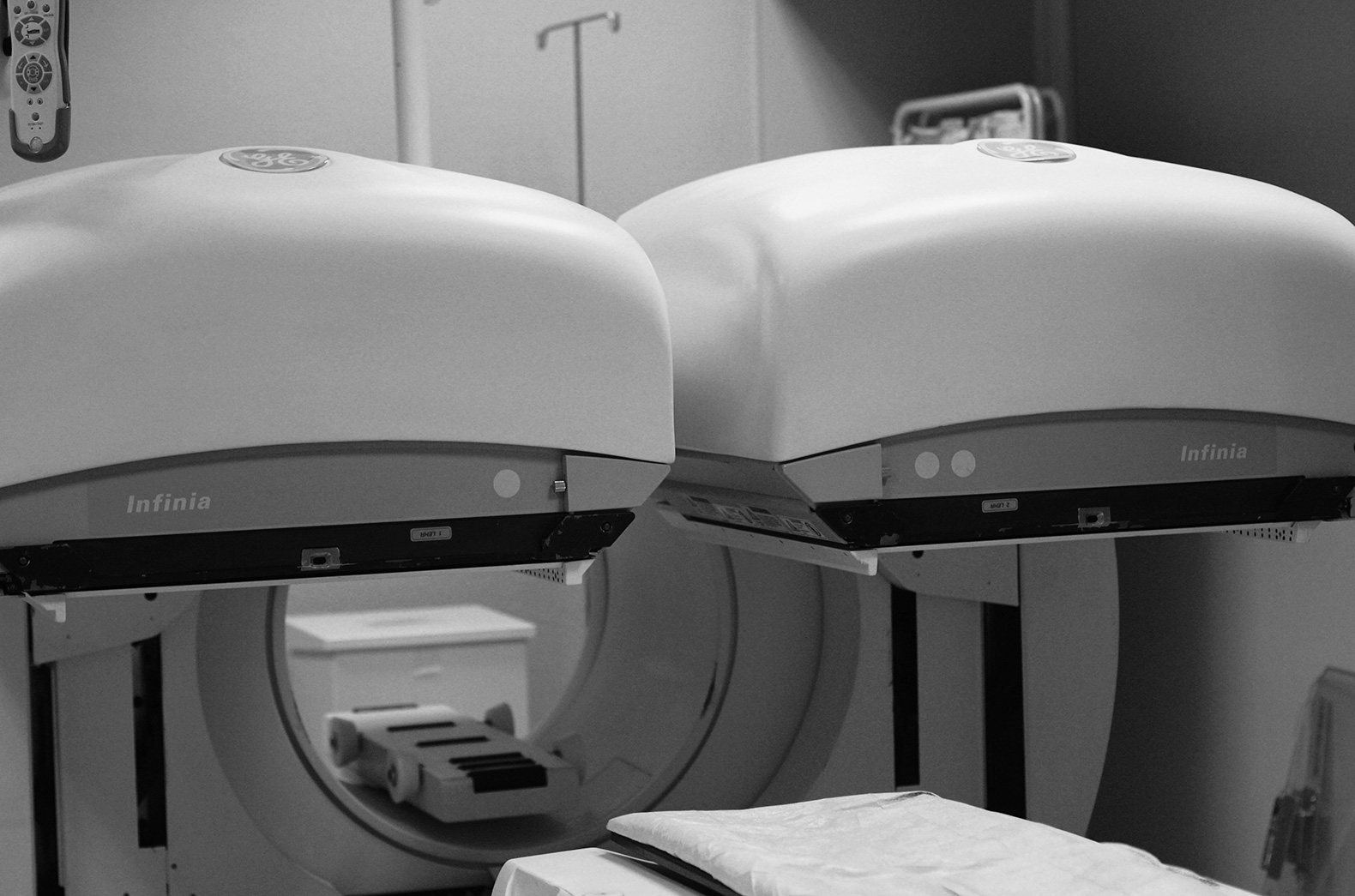
The rest study is initiated with the placement of an access way into an arm vein. It will be used for the administration of several drugs over the examination and will only be withdrawn after the exam completion. The radiopharmaceutical will then be administrated through the venous access, which doesn't cause any reaction. Later, the first images will be acquired in an equipment called Gamma CameraThe stress test is performed with cardiologist support and continuous monitoring of the electrocardiogram and blood pressure. Instead of the conventional exercise stress test (treadmill), a pharmacological stress test is performed. It consists on the administration of a drug that will exercise the heart according to the patient's weight. Any discomfort is transient. At the moment of maximum effort, patient will again be administered a tracer that will allow the heart’s visualization at that moment. Subsequently, a small snack will be offered. It is important for the quality of the results to insist on eating and drinking at this stage! After a new time interval, the patient will return to Gamma Camera for the last pictures, ending the test. 

3— Results 
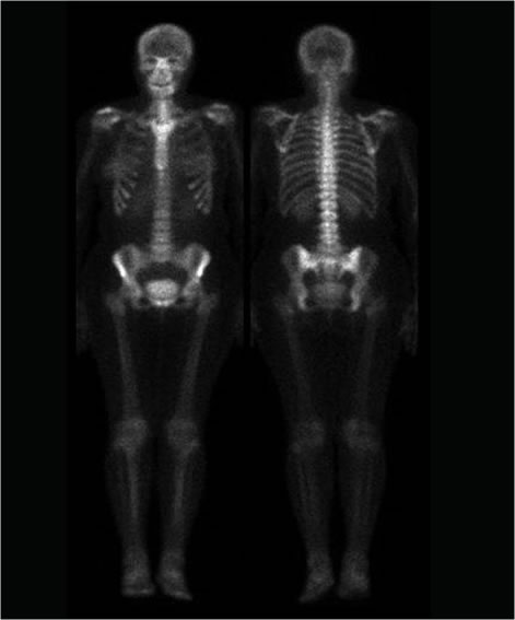
Our Nuclear Medicine Physician will subsequently analyse and compare the images of the two examinations (rest and stress). 
The examinations' report with images and the opinion of the cardiologist will be written to your doctor.

Notes

IMPORTANT

Documents to bring to the exam: 
— Doctor's Request;
— Social Security Card or other entity;
— Heart Exams;
— Previous Scintigraphy (if available); 
— List of current medication;
— Weight and height;
— Warn any accompanying person that the total duration of the exam is about 3,5 - 4 hours;
— The staff of ATOMEDICAL is at your complete disposal for any further information you may require.

Radionuclide Angiography (MUGA Scintigraphy)

Exam description

Notes

123I-MIBG Cardiac Scintigraphy

Exam description

Notes

Renogram (DTPA)

There are several reasons (congenital malformations, urinary tract infection, hypertension, renal dilatation, obstructive uropathy, etc.) that may give the physician (yours or your child's) the need to assess the functional status of the urinary tract. The required examinations will complete the necessary information for a complete diagnosis, helping to choose the best treatment, assessing the evolving maturity of the urinary tract or checking the results of a previous treatment.

The Renography will determine renal function on itself, i.e. how each kidney is to filter the blood and to excrete the urine.

Exam description

1 — Prior Preparation 
Proper hydration one hour before the exam is advised (250 to 500 ml, depending on age), but patient may urinate. For hypertension studies, your doctor may have to suspend or change your medication the week before the exam. 

2 — Procedure 
The patient is lying in the Gamma Camera, motionless for about 20 minutes. The tracer is administered through a small injection into a peripheral vein. In some cases, the study may require a second injection with some imaging extension (Diuretic Renography).

3 — Results 
Our Nuclear Medicine Physician will prepare the report for your doctor. Your doctor will give you all the necessary explanations about the results of this examination. 

Notes

Do not worry about the radiation dose received: it is usually lower than the corresponding radiological examinations. 

Diuretic Renography

Exam description

Notes

Captopril Renography

Exam description

Notes

Renal Scintigraphy

A renal scintigraphy ascertains the location of the kidneys, functioning renal mass and the presence of lesions (scarring, etc.).

Exam description

1 — Prior Preparation 
No preparation in advance is required. 

2 — Procedure 
The tracer is administrated on a small injection into a peripheral vein. The patient can then leave the Lab, returning one hour later. He's then laid in the Gamma Camera, motionless for about 10 minutes. 

3 — Results 
Our Nuclear Medicine Physician will prepare the report for your doctor. Your doctor will give you all the necessary explanations about the results of this examination. 

Notes

Do not worry about the radiation dose received: it is usually lower than the corresponding radiological examinations.

Direct Gamma Cystography

The cystography detects urine reflux from the bladder to the kidney.

Exam description

1 — Prior Preparation 
A negative urine culture is required, harvested no longer than 6 days before the exam (ideal 3-4 days)

Warning: You won't be able to make this exam if the prior analysis is not well made!
No other preparation is required. 


2 — Procedure 
The child is lying on the gamma camera. The doctor will disinfect the genital area and then place a very thin flexible tube into the bladder. The bladder is then filled with saline solution slowly by gravity. When the bladder is full, the child will urinate (on a previously placed diaper), thus ending the examination. It all takes around 30 minutes. 

3 — Results 
Our Nuclear Medicine Physician will prepare the report for your doctor. Your doctor will give you all the necessary explanations about the results of his examination. 

Notes

IMPORTANT
Bring the urine culture results, in the case of Gamma Cystography. 

ATTENTION
Without this analysis negative, you won't perform this examination!
Do not worry about the radiation dose received, it's usually lower than the corresponding radiological examinations.

Do not forget to bring:
— Your doctor’s request;
— Your social security card or other;
— Similar tests previously performed.

Indirect gamacistography

Exam description

Notes

Ventilation / Perfusion Lung Scan

Exam description

Notes

Perfusion Lung Scan

Exam description

Notes

Thyroid scintigraphy

Exam description

Notes

Parathyroid scintigraphy

Exam description

Notes

Gallium Scintigraphy

Exam description

Notes

Thallium Scintigraphy

Exam description

Notes

Adrenal Scintigraphy (iodocholesterol)

Exam description

Notes

Whole Body 123I-MIBG Scintigraphy

Exam description

Notes

Somatostatin receptors scintigraphy

Exam description

Notes

Sentinel lynph node Detection

Exam description

Notes

Whole body 131I scintigraphy

Exam description

Notes

Salivary glands scintigraphy

Exam description

Notes

Meckel’s Diverticulum Detection

Exam description

Notes

Gastro-Esophageal Reflex Detection

Exam description

Notes

Gastric Emptying Scintigraphy

Exam description

Notes

Esophageal Emptying Scintigraphy

Exam description

Notes

Lower Digestive hemorrhage Detection

Exam description

Notes

Labelled RBC hepatica Spect

Exam description

Notes

Hepatobiliary Scintigraphy

Exam description

Notes

Hepatosphenic Scintigraphy

Exam description

Notes

Brain SPECT (HMPAO or ECD)

Exam description

Notes

Brain SPECT (thallium)

Exam description

Notes

Loflupane and Brain SPECT (datascan)

Exam description

Notes

Thyroid Gland Ultrasound

Exam description

Notes

Salivary Glands Ultrasound

Exam description

Notes

Abdominal Ultrasound

Exam description

Notes

Iodine Therapy (Hyperthyroidism)

Exam description

Notes

Scrotal Scintigraphy

Exam description

Notes

Dacryoscintigraphy

Exam description

Notes

Antigranulocyte antibody whole body scintigraphy

Exam description

Notes